
Indian Paediatrics & Critical Care
Loading QBank Portal & Clinical Guidelines...
Loading QBank Portal & Clinical Guidelines...
Practice subtopic-wise clinical stations, one at a time, with a timed 4-minute timer & examiner checklist solutions.
Pick a subtopic from the left, review the module card, then hit Start Module.
0 Clinical Stations
STATION 5
Look at this acid-base nomogram and correctly label A to F.
Marks = 1*5 = 5
A. ………………………..
B. .………………………
C. .………………………
D. .………………………
E. .………………………
F.
………………………..
ANSWER KEY STATION 5
ANSWER
A. Metabolic acidosis
B. Acute respiratory alkalosis
C. Chronic respiratory acidosis
D. Acute respiratory acidosis
E. Metabolic alkalosis
F. Chronic respiratory alkalosis
 |
STATION 6
A 10 year old child came with C/O difficulty in breathing, coughing, nausea, vomiting, for 1 month, CBC showed eosinophilia, he was evaluated by chest X-Ray, and then advised CT chest?
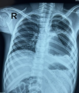
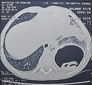
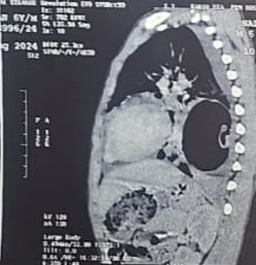
Marks è 0.5*5=2.5
Q 1) Identify the pathological sign in chest X-ray And CT chest? 0.5*2=1
Q 2) Clench the diagnosis and Write most common causative agent? 0.5*2=1
Q 3) Name the Most common organ involved by above agent other than lung? 0.5
Q 4) Write definitive treatment of choice in above condition? 1
Q 5) Write one precautionary measure during the definitive treatment of this condition? 0.5
Q 6) Write any 2 contraindications of pharmacotherapy? 0.5*2=1
ANSWER KEY STATION 6 A
Ans. 1) Chest X-Ray P/A view à large cavitary lesion in the left lower zone with internal air containing membranous structure capped by an air crescent—the ‘cumbo sign
A CT scan chest à cyst in left lower lobe of the lung containing a freely floating endocyst (the “water lily sign / Camelotte sign”)
Ans. 2) Echinococcus granulosus – most common
Note :- either any one below may give answer
1. Echinococcus granulosus: infections cause cystic echinococcosis, also called hydatidosis.
2. Echinococcus multilocularis: infections cause alveloar echinocccosis.
3. Echinococcus vogeli: infections cause polycystic echinococcosis.
4. Echinococcus oligarthrus: infections cause polycystic echinococcosis
Ans. 3) Primarily infect LIVER – most common then bypass to Lungs.
Ans. 4) Large cysts that are superficial and likely to rupture, infected cysts, cysts in vital anatomical locations, and cysts exerting substantial mass effect è SURGICAL INTERVENTION required è
· enucleation,
· pericystectomy,
· cystostomy with capitonnage,
· open aspiration, and lung resection. (lobectomy)
Ans. 5) Prevent intraoperative spillage by placing gauze soaked with a hypertonic saline solution (20%) or povidone-iodine solution
Ans. 6) Contraindication of pharmacotherapy è
Large cysts that are at risk of rupture
Inactive or calcified cysts
Bone marrow depression
Pregnancy, specifically the first trimester of pregnancy.