Indian Paediatrics & Critical Care

Loading QBank Portal & Clinical Guidelines...

Bedside Clinical Wisdom

Intensive Care Pearls

Daily high-yield PICU & NICU bedside pearls curated by Dr. Ajay Kumar Saini.

Bedside Pearls & Clinical Concepts (4)

High-yield clinical guidelines, drug dosing algorithms, and hemodynamics pearls.

💎 Newest published first
💎 Pearl 04

Difficult Intubation

✍️ Author: Dr. Ajay Kumar Saini

Definition of a physiologically difficult airway:

  • A physiologically difficult airway is where the patient has physiological characteristics that create a high chance of adverse events from the induction agents, ventilation or response to laryngoscopy, even though the laryngoscopy itself may be technically uncomplicated.

Contributing factors to the increased risk:

  • Risk of post-intubation hypoxia:
    • Reduced capacity for denitrogenation/preoxygenation (delirium, respiratory arrest)
    • Small FRC (obesity, pregnancy)
    • Increased demand (sepsis, thyrotoxicosis)
  • Risk of cardiovascular collapse
    • Any shock states, eg. hypovolemia, poor cardiac contractility, obstructive shock states
    • Reduced cardiovascular reserve, eg. unrevascularised coronary artery disease
    • Metabolic acidosis
    • Pro-arrhythmic state, eg. severe electrolyte derangement
    • Fragile aortic or cerebrovascular aneurysmal pathology
  • Risk of neurological injury
    • Raised ICP, hypertensive crisis

Optimisation strategies

  • Correct hypovolemia and acidosis
  • Pre-empt vasodilation and reduced cardiac output on induction with pre-intubation introduction of vasopressors and inotropes
  • Specifically look for, and target, RV failure (eg. with pulmonary vasodilators and inodilators)
  • Preoxygenate with NIV/HFNP
  • Use lower doses of cardiostable induction agents like ketamine
💎 DTaP Pedia Pearl 3

DTaP Pedia Pearl 3

✍️ Author: Dr. Ajay Kumar Saini


🖼️ Bedside Clinical Flowchart / Radiology Image:
Pearl Clinical Image
💎 Bedside Pearl

DFD

✍️ Author: Dr. Ajay Kumar Saini


💎 Bedside Pearl

IIN

✍️ Author: Dr. Ajay Kumar Saini

STATION 10

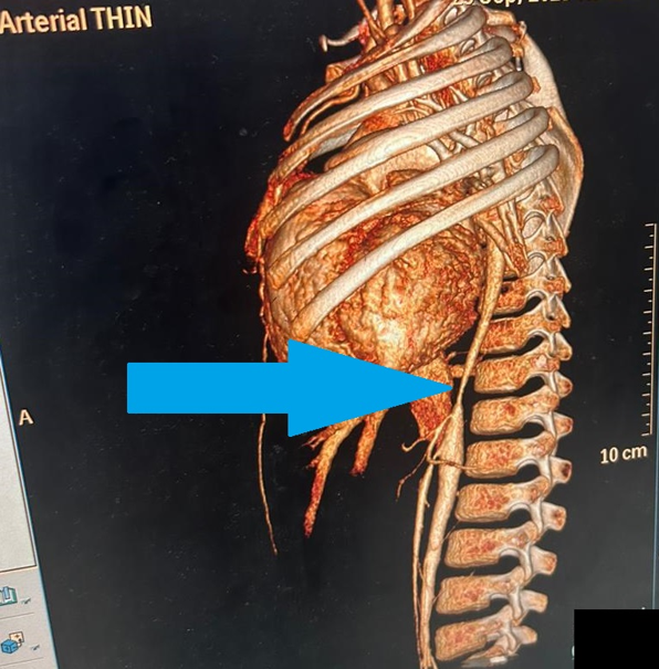
A previously healthy 6 year old female came with C/O fever for 2wk, and difficulty in breathing for last 3 days, on admission pt had tachycardia, tachypnoea with feeble femoral pulses and 2-D Echo show Dilated left ventricle with Ejection fraction <15%, BP in Upper limb was 134/96 while in Lower limb 72/54. A diagnostic imaging modality was done shown below in figure. Look at the figure and answer the following?

Marks

Q 1)     What is this diagnostic imaging modality ?                                                               1

Q 2)     What is the arrow showing in above depicted picture?                                            1

Q 3)     Identify the diagnosis ?                                                                                                1

Q 4)     Write any two differential diagnosis of above condition ?                             0.5*2=1

Q 5)     Write any one management option.                                                                             1


ANSWER KEY STATION 10

Ans. 1 )                  3-D reconstruction of CT AORTO-ANGIOGRAM

Ans. 2 )                  Stenosed segment of descending thoracic aorta

Ans. 3 )                  Takayasu arteritis – type II-III

Ans. 4 )                 (any two)

·        Aortic coarctation

·        Atherosclerosis

·        Behcet disease

·        Giant cell  arteritis

·        Ig-G4 related disease

·        Kawasaki disease

·        Rheumatoid arthritis

·        Sarcoidosis

Ans. 5 )                 (any one)

Corticosteroid (high dose iv steroid)

Immunosuppressive therapy (methotrexate)

Surgical revascularization

Arterial reconstruction